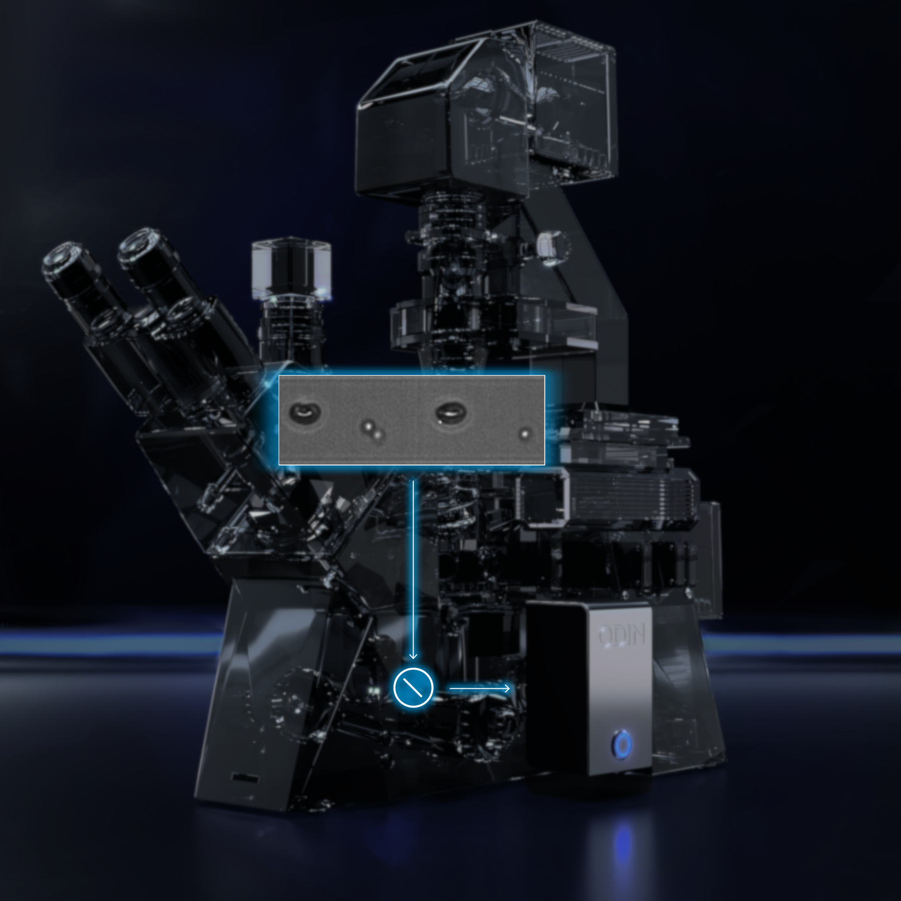
Image-Activated, Assay-Driven Cell Sorting
ODIN revolutionizes cell sorting with its ‘Seeing is Believing’ approach, transforming it from simple intensity peak analysis to a comprehensive, visual journey. It enables real-time, image-based insights, changing how researchers interact with and interpret experiments.
Product Overview
ODIN is a flexible solution that transforms any commercial microscope into a powerhouse for advanced cell sorting and analysis. This cutting-edge tool brings a “Seeing is Believing” ethos to cell sorting, transitioning from traditional intensity peak analysis to a rich, image-based examination. Completely label-free, eliminating the need for staining.
ODIN enables researchers to gain real-time insights into cellular interactions and behaviors, enhancing the understanding of cellular dynamics. Its dual sorting criteria, which incorporate both image-based and optionally fluorescence analysis, significantly reduce false positives and negatives, thereby improving the purity of sorted cell populations. With ODIN, the complexity of cellular functions becomes more accessible, opening new doors for in-depth research and the development of targeted therapies.
ODIN is versatile, supporting a wide range of applications from cell sorting and advanced microfluidics to microrheological measurements. See the table below for more details:
Image-Activated-Cell-Sorter (IACS) |
Our high-performance, imaging-based next generation cell sorter ODIN enables precise analysis and sorting of droplets, cells, spheroids, microorganisms, algae and other particles. |
Microfluidic Camera |
Utilize ODIN as a camera for your microfluidic experiments, benefiting from its high-performance software and SmartImaging feature to store only relevant data. Ideal for rare event detection and extended recording times. |
Droplet Generator |
Effortlessly generate, analyze, and sort droplets with the ODIN system, using its built-in feedback loop to control and manipulate droplet size. |
Particle Analyzer |
Analyze and sort any type of particle. |
Microrheology Sensor |
Perform user-friendly particle tracking with the VELOMIR ADD-ON, delivering state-of-the-art precision and speed. Continuously track and record up to 8 particles using a 15-parameter fitting procedure, achieving sub-pixel precision. |
Imaging Sensor |
High-performance camera with integrated analysis and feature extraction |
Frame Rate |
210 fps @ 1280×1024 px, up to 10000 fps @ 400×32 px, monochrome, 10bit color depth |
Sensitivity |
Wavelength range 300-1000 nm, 58% QE @ 550nm peak ; 62% QE @ 630nm; global shutter |
Readout Regions |
2 freely selectable, simultaneous readout regions, e.g. for two stage processes, immediate quality control or parallelization |
Latency |
Image capturing to analysis result <200 microseconds, control precision <10 nanoseconds |
Outputs |
6 flash outputs for light control; 2 trigger for sorting / device control (3.3V TTL) |
Interfacing |
C-mount; Ethernet; DVI-D; USB |
Software |
User-friendly and feature-rich graphical user-interface for observation, analysis, and control |
Analysis |
Real-time analysis of various detection parameters like size, velocity, position, granularity, circumference etc. |
Gating |
Freely configurable gating; graphical marking in scatter plot; arbitrary points polygon |
Triggers |
Freely programmable and independent triggers; latency down to 200µs (frame rate dependent) |
Storage Format |
RAW; AVI uncompressed; MJPEG; MP4; XLS; XLSX; CSV (infinite recording) |
Multi-Color |
Up to 4 simultaneous imaging settings by time multiplexing (e.g. bright field and three different fluorescent colors) |
Download the full ODIN Specification
Sorting Method |
Active Dielectrophoretic |
Sorting Efficiency, Selectivity, Purity |
Up to 99 % demonstrated |
Droplet Sizes |
10 µm to 1 mm diameter |
Detection Methods |
Bright field image; Phase contrast image; Fluorescence 408 nm image; Fluorescence 488 nm image; Fluorescence 532 nm image; Fluorescence 561 nm image; Fluorescence 638 nm image |
Detection Parameters |
Brightness/intensity; brightness contrast; circumference; area; surface roughness; colocalization; fluorescence colocalization; particle count; area contrast; fill factor; elongation; circularity; size; velocity; position; granularity; removal of small dirt particles; area of no contrast; contrast within particles; particles touching walls; particles touching image boundaries; particle position; particle velocity |
Modular Integration Options |
adaptable sorting techniques; adaptable microfluidic chips (no vendor lock in); adaptable microscope techniques; adaptable lasers; adaptable fluorescence filter sets; adaptable detection technologies; inline droplet generation or reinjection; incubation, temperature control; sterilization |
Quality Control |
In-line, multispectral image per particle pre & post sorting; all droplets recorded |
Sample Viability |
Further downstream usage of the sample; maximized cell viability |
Assay-based Sorting |
Yes |
Download the full ODIN Specification
(Singel) Cell Sorting
(Single) Cell Analysis
Cell Therapy
Drug Discovery
Drop-Seq
(RNA) Sequencing
Bacteria Analysis & Sorting
Algae Analysis & Sorting
Yeast Analysis & Sorting
Particle Analysis & Sorting
Sperm Analysis & Sorting
Spheroid Generation & Sorting
Droplet Generation, Manipulation, Analysis & Sorting
Microfluidic Camera Applications
Microrheological Experiments

Study Drug-Cell Interaction
Our innovative assay-driven sorting technology identifies cells based on specific drug interactions, offering direct insights into efficacy and mechanisms of action. In comparison to a conventional cell sorter, our system give you insights into the real-time behavior and direct response of a cell, not only morphological parameters. This breakthrough approach allows you to:
- Develop Patient-Specific Therapeutics: Tailor treatments to individual patient needs for maximum effectiveness.
- Enhance Effectiveness: Improve the precision and impact of therapeutic interventions.
- Accelerate Development: Speed up the process of bringing new treatments to market.
Enhance Microbiological Insights
Our advanced technology enables the direct observation of individual responses from various microorganisms and cells, even without staining. Our systems utilize stand-alone imaging-based, stand-alone fluorescence-based, or a combination of both methods. We don’t just use images for quality control; we use them for actual sorting decisions. This allows us to sort based on specific positions, interactions between different organisms under investigation, or their individual responses. Additionally, our technology offers high sorting speed, maximized cell viability, and full sterility. Explore the following applications, among many others:
- Isolate High-Performers: Identify high-performing drug-producing bacteria, food-producing microorganisms, or cells.
- Analyze Behavior: Study the individual behavior bacteria, algae, yeast, or other cells by measuring critical parameters.


Advance Biological Research
Our sophisticated technology enables precise analysis and sorting of cells, microorganisms, and other particles, facilitating crucial research in areas such as antibiotic resistance and pathogen identification. It can enhance your sequencing and supports you in various steps of your individual process.
- Sort Based on Individual Configurations: Look for co-localization, shape, or a defined number of specific cells.
- Run Full Assays: Utilize droplet microfluidics as micro-confinements to run assays with low sample consumption and high precision
Pioneer Personalized Medicine
Our cutting-edge technology allows you to sort based on individual cellular responses, enabling the isolation of high-performing killer cells or other cells with precision. This advanced capability allows researchers to gain profound insights into cellular functions under a variety of assay conditions, providing a deeper understanding of how cells behave and interact in different environments.
- Tailor Treatment: Meet the unique needs of individual patients, ensuring personalized and effective therapeutic strategies.
- Instant, Visual Quality Control: You can monitor and assess the quality of your experiments in real-time, making adjustments as needed to optimize results.
Experience the ease and efficiency of controlling your experiments with our user-friendly interface, designed to streamline your workflow and enhance productivity, paving the way for groundbreaking discoveries and innovations.

Comprehensive Imaging: Capture droplets in real-time while reducing storage demands by up to 95%.
Enhanced Visualization: Experience multi-colored and fluorescent imaging with up to 4 parallel modes.
Crystal Clear Images: Benefit from sharp imaging thanks to our ultra-short, pulsed illumination.
Instant Quality Control: Fully integrated real-time data analysis eliminates extensive post-processing, even at imaging rates of up to 10,000 frames per second.
User-Friendly Interface: Gate your sample within a couple of seconds with our intuitive user interface.
Integrated Analysis: Combines morphological and fluorescent marker analysis in one step.
Sterility: Ensures high cell viability and maintains sterility with enclosed channels.
Speed and Efficiency: Boost efficiency and efficacy of encapsulation to an unmatched level.